Evolution des caractères crâniens et endocrâniens chez les Afrotheria (Mammalia) et phylogénie du groupe
Anatomie comparée du labyrinthe osseux des Bibymalagasia, des Tubulidentata et des Tenrecoidea Ce sous-chapitre correspond à l’article suivant: Benoit J., Lehmann T., Vatter M., Lebrun R., Merigeaud S., Costeur L., Tabuce R. (In prep.) Comparative anatomy and three dimensional geometric-morphometric study of the bony labyrinth of Bibymalagasia (Mammalia, Afrotheria).
Abstract
Plesiorycteropus (Malagasy aardvarks) is the sole genus belonging to an extinct mammalian order that lived in Madagascar until recent time, the Bibymalagasia. Their systematic and phylogenetic position is controversial. Firstly, given that Plesiorycteropus morphologically resembles an aardvark (Tubulidentata), many authors have related them to Tubulidentata. Secondly, a more recent molecular analysis has proposed that they belong to Tenrecoidea, along with Tenrecidae (tenrecs and otter-shrews) and Chrysochloridae (golden moles). This context of competing phylogenetic hypotheses is stimulating for the investigation of new characters for phylogenetic inferences. Here we used the non-invasive methods of micro-CT scanning, digital reconstruction and three-dimensional geometricmorphometric analysis of shape to investigate the morphology of the bony labyrinth (osseous inner ear) of the holotype skulls of both known species of Bibymalagasia: Plesiorycteropus madagascariensis and P. germainepetterae. Firstly, we find that the bony labyrinth of Bibymalagasia is distinctive from that of other afrotherians examined, supporting their ordinal distinctiveness. Secondly, the PCA and the Neighbour Joining tree on labyrinthine shape data show that the bony labyrinths of both Plesiorycteropus more closely resemble those of tubulidentates than to that of any other afrotherian. If this similarity reflects phylogeny, then it 37/468 Bibymalagasia, Tubulidentata et Tenrecoidea could support a sister-group relationship between Bibymalagasia and Tubulidentata. Conversely, if it is homoplastic, there is one character that could support the clade uniting Bibymalagasia and Tenrecoidea: the shared absence of a secondary common crus (partial fusion of the lateral and posterior semicircular canals). Indeed, this character is primitively present in extant and extinct Tubulidentata investigated. We also find that when compared to living tubulidentates and tenrecoids, the bony labyrinth of both species of Plesiorycteropus reveals a certain degree of variation that rather suggests intra- instead of inter-specific variability.
Introduction and systematic background
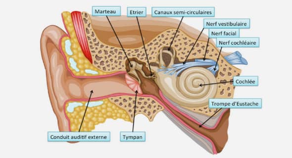
Bibymalagasia is an order of Malagasy mammals represented by the sole sub-fossil genus Plesiorycteropus (Patterson, 1975; MacPhee, 1994). This genus lived approximately 2000 years BP (Burney et al., 2004). It is often called the ‘Malagasy aardvark’ because of some morphological similarities it shares with true African aardvarks (genus Orycteropus) (e.g. Lamberton, 1946; Patterson, 1975; Wible, 2012). In fact the generic name Plesiorycteropus, as it was defined by Filhol (1895), means “near Orycteropus”. Nowadays, the African species Orycteropus afer is the sole representative of the Tubulidentata, but this order was much more diversified and widespread in the past, including occurrences in Pakistan, Middle East, Greece and France (Tabuce et al., 2008; Lehmann, 2006, 2009). Orycteropodidae represents the sole family of the order Tubulidentata since Plesiorycteropodidae (including Plesiorycteropus) were assigned to their own order by MacPhee (1994). For this author, most similarities shared by Orycteropodidae and Plesiorycteropodidae were likely acquired convergently due to their fossorial habits and adaptation to digging (or climbing) and ant-eating. Therefore he erected the new mammalian 38/468 Chapitre 1 order Bibymalagasia in « recognition of the distinctiveness of Plesiorycteropus » (MacPhee, 1994: 201). The systematic and phylogenetic positioning of Plesioryctropodidae among mammals is a long standing debate. Indeed, the evolutionary history of Plesioryctropodidae is all the more complicated by the fact that they display a mixture of both apomorphic and plesiomorphic characters (see MacPhee, 1994) which testifies to the great antiquity of the lineage that led to the Quaternary genus Plesiorycteropus. Their origin could even be dating back to the Eocene epoch or even earlier, at the time when their first representatives landed on Madagascar (Patterson, 1975). Since the work of MacPhee, many subsequent phylogenetic analyses have however supported a sister-taxa relationship between Bibymalagasia and Tubulidentata (Asher et al., 2003: fig. 2; Horovitz, 2004; Holroyd and Mussell, 2005; Asher et al., 2005: fig. 9A; Asher, 2007: fig. 2), while some others branch it elsewhere among Afrotheria (Asher et al., 2005: fig. 9B) or elsewhere in the mammalian tree (Asher et al., 2003: fig. 5B; Asher, 2007: fig. 1). Consequently, many recent workers preferred to avoid debating possible affinities and accepted MacPhee´s hypothesis that Bibymalagasia is a distinct mammalian order of unclear affinities (e.g. McKenna and Bell, 1997; Lehmann, 2009). In parallel, recent molecular studies have greatly increased confidence in placental phylogeny and gave support to a new clade nesting Tubulidentata with some other African placental mammals (e.g. elephants, hyraxes and elephant-shrews) (Stanhope et al., 1998; Springer et al., 2004). Since the discovery of this clade, called Afrotheria, many recent morphological, combined morphological and molecular, and supertree analyses have supported the inclusion of Bibymalagasia into Afrotheria (Asher et al., 2003: fig. 2; Asher et al., 2005: fig. 9A; Beck et al., 2006; Asher, 2007: fig. 2). Consequently the inclusion of Bibymalagasia into Afrotheria has been generally accepted (Kemp, 2005; Werdelin and Sanders, 2010). Recently, a crucial work based on molecular sequence obtained from fossil 39/468 Bibymalagasia, Tubulidentata et Tenrecoidea bone collagen of Bibymalagasia has deeply clustered Plesiorycteropus into Afrotheria and Tenrecoidea (Chrysochloridae + Tenrecidae), as the sister group of Tenrecidae (Buckley, 2013). Hence, this recent study definitely shows that Bibymalagasia belong to Afrotheria. More disputable is the nesting of Plesiorycteropus within Tenrecoidea, because the interordinal relationship in the tree obtained by Buckley (2013: fig. 4) is not consistent with those elsewhere proposed (e.g. Stanhope et al., 1998; Springer et al., 2004). The most striking examples are the branching of Orycteropus within Paenungulata as the sister-group of Hyracoidea, or that of the Macroscelidea Petrodromus at the base of the afrotherian tree (Buckley, 2013: fig. 4). Interestingly, a clade uniting Tenrecoidea and Plesiorycteropus, as figured in Asher et al. (2005: fig. 9B), would be supported by three homoplastic morphological characters (optic foramen smaller than the sphenorbital fissure, humeral head standing superior to the greater tuberosity, reduced pubic symphisis). MacPhee (1994) did not use Tenrecoidea in his matrix, but he used Tenrec ecaudatus as chief taxon to code the Soricomorpha. None of his analyses, whatever the assumption, support a clade clustering Plesiorycteropus with Soricomorpha (MacPhee, 1994). On the contrary, the parsimony analysis of his full datamatrix supports the clade (Plesiorycteropus, Orycteropodidae) with three characters: the reappearance of the percranial canal, the presence of thoracolumbar transarcual canals and the presence of a lateral process of the ischium. These synapomorphies were latter supplemented by six other characters (loss of the subarcuata fossa, expanded trochlear facet of the radius, presence of more than five sacral vertebrae, anterior process of the pubis absent, presence of a cotylar fossa of the astragalus and posteromedial flange of the astragalus present) in the works of Asher (2005, 2007) and Asher et al. (2003, 2005). Unfortunately, all these characters (except the presence of the lateral process of the ischium) are again homoplastic within afrotherians or placentals, and their presence in Tubulidentata and Bibymalagasia could be the result of their convergent adaptation to fossorial habits and 40/468 Chapitre 1 ant-eating (robust limbs for digging (or climbing) and slender masticatory apparatus) (MacPhee, 1994). Wible (2012) pointed out some similarities of the middle ear between Orycteropus and Plesiorycteropus, but whether they constitute true shared derived trait remains to be addressed. In a recent paper, it has been proposed that Bibymalagasia and Tubulidentata could share the presence of a unique morphological trait on the neopallium (Benoit et al., 2013e1 ). Indeed, using X-ray tomography and microtomography (CT-scan) to investigate the endocranial casts of afrotherian mammals, these authors found that Bibymalagasia and Tubulidentata were the only species that share a contact between the praesylvian sulcus and the lateral sulcus of the neopallium . Being absent in other mammals, and even in fossorial species, this character could constitute a robust synapomorphy of Tubulidentata and Bibymalagasia. It chiefly demonstrates the importance of new characters for systematic and phylogenetic discussions and the rich potential of CT-scan surveys of fossils to provide useful new characters. CT-scanning is a non-destructive and non-invasive technique that allows the study of previously hardly accessible structures such as the endocranial cast and the bony labyrinth (osseous inner ear) of extant and extinct species. As a consequence, the number of CT-scan assisted studies of the bony labyrinth of vertebrate has dramatically increased during last two decades (e.g. Spoor and Zonneveld, 1995; Spoor et al., 2002; Lebrun et al., 2010; Orliac et al., 2012a; Ekdale, 2013). Dental anatomy has been and continues to be one of the major sources of character to assess mammal phylogeny and relationships. It is also true in Tubulidentata in which the columnar, rootless and hypselodont teeth, made of hundreds of tubular prisms of dentine, has inspired their ordinal name (Holroyd and Mussel, 2005). However, the use of tooth morphology to determine tubulidentates relationship is limited by their simple dental pattern, displaying neither cusps nor crests (Lehmann, 2009). Moreover, dental characters are 1 See chapter 5 41/468 Bibymalagasia, Tubulidentata et Tenrecoidea not available for Bibymalagasia since Plesiorycteropus is known by post-cranial and basicranial material only and no maxillaries, mandibles, nor isolated teeth have been found yet (MacPhee, 1994; Lehmann, 2009). Fortunately, at least two specimens of Plesiorycteropus preserved their ear region (MNHN-MAD 327 and MNHN-MAD 328). Consequently, in the absence of further new dental material documenting the teeth morphology in Plesiorycteropus, to study the bony labyrinth of Plesioryctorepus seems the best way to bring new and previously unpublished characters to the debate on the systematic position of Bibymalagasia.
Material and methods
Material
The holotypes of both species of Plesiorycteropus, housed at the Muséum National d´Histoire Naturelle (MNHN), Paris (France), were scanned. They consist of MNHN-MAD 327 (P. germainepetterae) and MNHN-MAD 328 (P. madagascariensis) (Fig. 1). Scans were done at the AST-RX µCt-scanning station v tome xL 240 (Paris, France) with a voxel size of 35.7µm (MAD 327) and 39.7µm (MAD 328). Segmentation and 3D reconstructions of their bony labyrinth were done using Avizo 6.3 (VSG) software. The bony labyrinths of these specimens were compared to those of four species of Tenrecoidea (three Tenrecidae, one Chrysochloridae), to the extant aardvark Orycteropus afer, and to two species of fossil aardvarks, Amphiorycteropus gaudryii (type species of the genus Amphiorycteropus Lehmann, 2009) from the upper Miocene of the Island of Samos (Greece) and Amphiorycteropus depereti (NMB Rss. 55, the holotype of the species) from the Pliocene of Perpignan (France) (see Annexe 1.1 for a list). 42/468 Chapitre 1 Figure 1. CT-reconstruction of the skulls and bony labyrinth of holotypes of Plesiorycteropus germainepetterae (MNHN MAD-327) (A) and P. madagascariensis (MNHN MAD 328) (B) in lateral view.
Measurement protocol.
Measurements of all specimens’ bony labyrinth (Table 1) were performed with Avizo 6.3. A summary of the measurement protocol is provided in figure 2. The length of the bony labyrinth is the greatest length of the bony labyrinth from the ventral most border of the cochlear canal to the dorsal most point of the posterior semicircular canal when viewed in posterior view (Fig. 2A). The stapedial ratio (Segall, 1970) is the quotient of the length and the width of the fenestra vestibuli (Fig. 2B). A stapedial ratio exceeding 1.6 corresponds to a rather oval fenestra vestibuli, while a value below 1.6 corresponds to a rather rounded aperture (Benoit et al., 2013b). The number of turns of the cochlear canal corresponds to the number of turns completed by the canal from its apex to the end of the primary bony lamina (Fig. 2C), in degrees (Geisler and Luo, 1996). The cochlear 43/468 Bibymalagasia, Tubulidentata et Tenrecoidea length was measured along the spiral of the cochlear canal taken from its apex to the most proximal end of the primary bony lamina (Fig. 2C). The cochlear ratio, or aspect ratio of the cochlear canal (see Ekdale, 2009), corresponds to the quotient between the width and the height of the cochlear canal when viewed in profile (Fig. 2D). The diameter of the cochlear canal corresponds to the width between the posterior most point of the centre of the lumen of the cochlear canal and the anterior-most one (Fig. 2C). The angles between the planes of two semicircular canals are measured when the plane of both semicircular canals are perpendicular to the field of view (see Ekdale, 2009) (Fig. 2E, F, G). The ventral expansion of the posterior semicircular canal (V.e.p) (equivalent to the sagittal labyrinthic index of Spoor and Zonneveld (1995)) is measured when the plane of the lateral semicircular canal is parallel to the horizon (Fig. 2B). After Spoor and Zonneveld (1995) this measure is the ratio of the distances between the level of the ventral-most point of the lumen of the posterior semicircular canal and the plane of the lateral semicircular canal, and that between the level of the dorsal-most point of the posterior canal and the plane of the lateral semicircular canal. The dorsal expansion of the anterior semicircular canal (D.e.a) (equivalent to the extension of the anterior semicircular canal projecting to the dorsal-most point of the posterior semicircular canal of Schmelzle et al., 2007) is measured when the plane of the lateral semicircular canal is parallel to the horizon and the plane of the posterior semicircular canal is perpendicular to the field of view (Fig. 2B). This measure is the ratio of the distances between the level of the dorsal-most point of the lumen of the posterior semicircular canal and the level of the dorsalmost point of the lumen of the anterior one, and the length between the dorsal-most point of the lumen of the posterior semicircular canal and the ventral-most point of the anterior semicircular canal and the surface of the vestibule. The radius of a semicircular canal is half.
Evolution des caractères crâniens et endocrâniens chez les Afrotheria (Mammalia) et phylogénie du groupe |