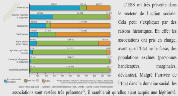
Living actinopterygian
Dorsal and anal fins (Fig. 1 ) developmental sequences were obtained from embryojuvenile rainbow trout (Oncorhynchus mykiss). Examined specimens range from 8 days pre-hatching to 100 days post hatching (dph). Larva-juvenile were reared in a swimming channel (see Grünbaum et al. 2008 for detail on rearing system) under constant water velocity (004 cm/s) in 2005 (see Chu 2007 for rearing conditions). Specimens were sampled every day from 8 days pre-hatching to 34 dph, every other day from 34 to 80 dph, and every four days up to 100 dph. Samples were fixed in neutral buffered formalin for 48h, and then preserved in 70% ethanol. Two series of cleared-and-double stained (Alizarin red S for bon es and Alcian blue for cartilages: Dingerkus and Uhler 1977; Potthoff 1984) specimens have been used in this study. Pre-hatching specimens were removed from their egg capsule prior to be cleared and stained solely with Alcian blue.
Digital photographs were taken before staining and 5-10 days after staining to avoid interpretive errors owing to destaining. The first series includes one specimen for each sampling day. The second series was used to cope with staining problems (Redfern et al. 2007); additional specimens from 0 to 24 dph were stained. A total of 86 specimens have been examined to reconstruct developmental sequences. Ali observations were made under a Leica MZ16A stereodissecting mIcroscope equipped with a digital camera. Standard length (SL) was measured prior to staining with Northern Eclipse Software (Version 6.0, Empix Imaging Inc. , Ontario, Canada). Since SL and dph are highly correlated (r2 = 0.952; P < 0.001) and SL is recognized as a better proxy for morphological development in fishes (Fuiman et al. 1998; Faustino and Power 1999), SL was used for aIl statistical analyses.
Co ding was based on color uptake (i.e. , cartilages are blue and bones are red) and color intensity (i.e., first structures to take col or are darker). Developmental states for radiais are: (1) present (ceU condensation without stain uptake), (2) cartilaginous (blue), and (3) ossified (red). Developmental states for lepidotrichia are: (1) present, (2) ossified (red), (3) segmented (number of segments per lepidotrichium), and (4) bifurcated (position of the bifurcation). Because the number of seriai e1ements (i.e., radiais and lepidotrichia) varies among individuals, positional homologies and numbering of elements (e.g., dorsal radiais 1-14, dorsal lepidotrichia 1-17, anal radiais 1-14, anal lepidotrichia 1-16) were inferred a posteriori by lining up aU specimens with the third radial (variability being more important in peripheral areas) and by comparing similarities between sequences of similar sized-specimens. Furthermore, myomere counts (from head to tail) were used in the earliest larval stages as a topographical criterion to identify the first proximal radiaIs to differentiate. The dorsal fin is positioned at the level of myomeres 21-32, whereas the anal fin is positioned at the 1evel of myomeres 40-50. The ske1etal e1ements were numbered following their order from anterior (1) to posterior (up to 17).
Logistical regressions were used to estimate the standard length (SL) at which 50% (SLso) of the specimens have reached the developmental state of interest (i.e., cartilaginous, ossified, segmented and bifurcated) for each element. Values of SLso were subsequently used to order and reconstruct developmental sequences among seriaI elements within a fin. Significance of the logistical regressions was tested using the G2 statistic (Quinn and Keough 2002). The significance level used to interpret a regression for a given element was ca\culated using the Bonferroni correction: the collective significance level of 0.05 was divided by the number of elements to get the nominal significance level for each regression. Statistical analyses were performed using SYST A T (Version 11.00.0 1, SYSTAT Software Inc., 2004, Richmond, Califomia). In order to validate the DAFPM and the EEM, Spearman rank correlation coefficients were used to describe the relation between corresponding sequences in the dorsal and anal fins, and in the exoskeleton and endoske1eton. Only the elements for which the logistical model was significant were considered for Spearman correlations. The significance level used was 0.05.
Living actinopterygian
The 86 specimens of 0. mykiss examined have a SL that ranges from 10.1 to 32.7 mm. The number of proximal radiaIs is variable in the dorsal and anal fins among indi viduals, ranging from 6 to 15 radiaIs in the dorsal fin and from 3 to 14 radiaIs in the anal fin. Linear regressions were used to verify the cause of this variability by estimating the relation between the number of elements (radial and lepidotrichia) and SL. This variability is mostly due to ontogenetic changes; there is a significant positive correlation between SL and the number of radiaIs in the dorsal (rs = 0.682; P < 0.001) and the anal (rs = 0.635 ; p < 0.001) fins. The unexplained portion of the variability is probably due to differences among individuals. Each proximal radial is usually associated with one distal radial. The only exception concerns the anteriormost proximal radial where the distal radial is frequently missing; lepidotrichia are thus articulated directly with the proximal radial. Ontogenetic changes are aiso good explanations for the variability in the number of lepidotrichia in the dorsal (rs = 0.817; p < 0.001) and anal (rs = 0.857; p < 0.001) fins. There are up to 18 lepidotrichia in the dorsal fin and up to 16 in the anal fin. There is al: 1 relationship between the number of radiais and lepidotrichia except for the first and last radiais: three to four lepidotrichia are associated with the first radial and two lepidotrichia are associated with the last radial (Fig. 1). Eight events were studied in the development of the dorsal and anal fins (Table 2). Their arder of initiation is: (1) differentiation of proximal radiais, (2) chondrification of proximal radiaIs, (3) differentiation of lepidotrichia, (4) chondrification of distal radiais, (5) segmentation of lepidotrichia, (6) ossification of lepidotrichia, (7) bifurcation of lepidotrichia, and (8) ossification of proximal radiais. After initiation, these developmental events proceed simultaneously.
The differentiation of proximal radiais begins 5 days before hatching in specimens reaching 10 mm in SL. The first proximal radiais to differentiate are radiaIs 4-9 in the dorsal fin and radiais 4-10 in the anal fin. Radial differentiation is quickly followed by chondrification. In fact, only three specimens (pre-hatched and 0 dph) show cell condensations in one or both fin s prior to the beginning of chondrification. Consequently, the differentiation of proximal radiais was not analysed statistically owing to the lack of specimens showing this state. The first cartilaginous proximal radiais for both fins form in a 12 mm long specimen. The first proximal radiais to chondrify are radiais 4-9 in the dorsal fin and radi ais 6-8 in the anal fin. The sequences of chondrification of the proximal radiaIs are highly congruent between both fins (rs = 0.919; p < 0.001 ; Table 3), but a slight timing divergence occurs posterior to radial 8 (Fig. 3A). The formation of actinotrichia precedes the formation of lepidotrichia, which differentiate proximo-distally . The differentiation of lepidotrichia begins with only a slight de1ay after the differentiation of the first proximal radial in a 14 mm long specimen (Table 3). The first lepidotrichia to form are lepidotrichia 7-11 in the dorsal fin and lepidotrichia 6- 9 in the anal fin (Table 2). The pattern of differentiation of the lepidotrichia is highly congruent between both fins (rs = 0.860; p < 0.001 ; Table 3, Fig. 3B), but a timing divergence is shown posteriori y to lepidotrichia Il (Fig. 3B). The pattern of differentiation is congruent between the endoskeleton and the exoskeleton; the first lepidotrichia to form are supported by the first proximal radiais to appear.
Actinistia Two actinistian species have been examined: (1) the Late Devonian Miguashaia bureaui and the Late Carboniferous Rhabdoderma exiguum. M. bureaui is considered as an actinistian basal taxon (Cloutier 1991, 1996a), whereas R. exiguum is a typical Carboniferous actinistian. Both species where first described from juvenile specimens; but while M. bureaui is known from few specimens, with a great size range (Cloutier 1996a), R. exiguum (Eastman 1902, 1903) is essentially known from small juvenile specimens (Schultze 1972, 1985). There are 27-28 lepidotrichia in the second dorsal fin and ca. 25 in the anal fin of M. bureaui. The longest lepidotrichia are in the anterior part of the fin at the level of lepidotrichia 4-6 in the dorsal fin and at the level of lepidotrichia 7-8 in the anal fin (MHNM 06-41; Fig. 9). This pattern is not seen in the juveni1e specimen (ULQ 120b; Fig. 8) because the distal part of both median fins is not preserved. In the juvenile specimen ULQ 120 (Total length (TL): 72 mm), the basal proximal segments are 2.8 mm long in the dorsal fin and 2.1 in the anal fin (Fig. 8). In specimen MHNM 06-41 (TL: 195 mm; Fig. 9), these proximal segments are longer; the basal segments reach 3.3 mm in the dorsal fin and 7.2 mm in the anal fin. Moreover, the length of the basal segments in both fins reach 9-10 mm in specimen MHNM 06-494 (TL: 375 mm). In specimen MHNM 06-41, the first segment distal to the basal element of lepidotrichium Il seems to be half-merged with the basal e1ement (Fig. 9). This observation is interpreted as a merging of proximal segments; thus explaining the 1ength increase of the basal segment during growth. Whi1e the length of the basal elements increases, the size of the distal elements remains unchanged at 2 mm. However, while the total length of the distal segments does not increase, sorne important shape changes occur. In the juvenile specimen of M. bureaui (ULQ 120b; Fig. 8), lepidotrichial segments have a thin-rod shape with sorne segments showing small anteroproximal and posterodistal extensions. This characteristic interlocking pattern intensifies during growth and becomes more apparent in larger specimens [MHNM 06-41 (Fig. 9), MHNM 06-494 (Fig. 10)].
REMERCIEMENTS |