Antimicrobial activities of Dilobeia thouarsii Roemer and Schulte, a traditional medicinal plant
Traditional medicine is an important component of the health care 52 system in Madagascar and a large number of plants remain to be 53 studied, including Dilobeia thouarsii, a tree that belongs to the family 54 Proteaceae and is endemic to Madagascar (Boiteau, 1986). This 55 species is widely distributed in the Central, Eastern, South-Eastern 56 regions and in the high Matsiatra Fianarantsoa in Madagascar 57 (Bosser and Rabevohitra, 1991) and is known by the common 58 names of Vivaona, Hazontavolo and Tavolohazo (Rabesa, 1986). In 59 southern Madagascar, decoctions of the leaves and bark of D. thouarsii 60 are used for abortion, or as an anthelmintic, or a diuretic (Beaujard, 61 1988; Rabesa, 1986). Concerning the East coast of Madagascar 62 (Mandraka region), our ethnobotanical investigations confirmed 63 the use of the leaves in traditional medicine to treat bacterial skin 64 infections and wounds (Razafintsalama, 2012). In vitro assays have shown that phenolic compounds are often belonging to this family display antimicrobial activities against different 73 microorganisms. L. hirsuta, which is used in traditional medicine 74 in Chile, is active against the pathogenic fungus Candida albicans 75 (MIC = 8 μg/ml) (Simonsen et al., 2006). A phenolic glycoside ester 76 isolated from the New Zeland tree T. toru is active against Pseudomonas 77 aeruginosa, Escherichia coli and Bacillus subtilis (Perry and Brennan, 78 1997). A glycoside compound isolated from Persoonia linearis × pinifolia, 79 a cross hybrid of P. pinifolia and P. linearis, displays antimicrobial activity 80 against E. coli and Phytophthora cinnamoni (MacLeod et al., 1997). An 81 extract made from leaves of Protea simplex, a plant used in South Africa 82 against human dysentery and diarrhea, provides good antimicrobial Contents lists available at SciVerse ScienceDirect South African Journal of Botany journal homepage: www.elsevier.com/locate/sajb Please cite this article as: Razafintsalama, V., et al., Antimicrobial activities of Dilobeia thouarsii Roemer and Schulte, a traditional medicinal plant from Madagascar, South African Journal of Botany (2013), activities against E. coli, Staphylococcus aureus, B. subtilis and C. albicans 85 (Fawole et al., 2009). 86 To the best of our knowledge, no report has been published on the 87 chemical composition and the biological activities of D. thouarsii 88 (Bosser and Rabevohitra, 1991). In the present study, we investigated 89 the antibacterial activity of D. thouarsii and identified bioactive 90 compounds in order to provide a scientific basis for its traditional use, 91 and to characterize the potential of this medicinal plant in Madagascar. 92 Bioassay fractionation enabled isolation of two phenolic compounds 93 that were identified on the basis of spectroscopic data including 1D 94 NMR and mass spectrometry (MS).
Material and methods
Plant material
The leaves of D. thouarsii were harvested in Mandraka region, in the 98 eastern part of Madagascar, 70 km from Antananarivo. Leaves were col99 lected in April 2008. The plant was identified by Dr. Rabarison Harison 100 from the Botany Department of Antananarivo Faculty of Sciences. 101 Reference specimens (HERB/DBEV/4708) were deposited in the 102 herbarium of the same department of the University of Antananarivo.
Extraction of D. thouarsii leaves
Plant materials were dried at room temperature and ground to a fine 105 powder. The obtained powder (100 g) was extracted successively 106 through a maceration process using 500 ml × 6 of solvents of increas107 ing polarity (hexane, ethyl acetate and methanol). Each combined 108 extract was evaporated under reduced pressure to yield crude hexane 109 extract (0.7 g), EtOAc extract (5 g), and MeOH extract (10 g), respec110 tively. Extracts were stored at room temperature until use. 111 2.3. Bioassay-guided extract 112 Part of the ethyl acetate extract (1.5 g) was subjected to flash 113 chromatography on a silica gel 60 (10–40 μ) column (CC) eluted 114 with 0–100% gradient of EtOAc in hexane followed by MeOH in 115 EtOAc. Fourteen 100 ml fractions were collected: Hex–EtOAc 80:20 116 (1–4), Hex–EtOAc 40:60 (5–8), Hex–EtOAc 20:80 (9–12), EtOAc 117 (13), and MeOH (14). On the basis of the analytic TLC, and according 118 to the antimicrobial assay, similar active fractions 5–8 (0.15 g) 119 were combined and rechromatographed on the same support using 120 the same solvent system. Fourteen new fractions were obtained, 121 but only two displayed antibacterial activity. Each active fraction 122 was treated with 75% ethanol and concentrated to yield compounds 123 1 (100 mg) and 2 (40 mg). The antimicrobial activity of these com124 pounds was evaluated on Gram-positive and Gram-negative bacteria.
Antimicrobial assays
Microorganism strains
Four Gram-positive (Bacillus cereus LMG 6910, Bacillus megaterium 128 LMG 7127, S. aureus ATTC 25920, Enterococcus faecalis ATTC 29212) 129 and six Gram-negative bacteria (Vibrio harveyi ATCC 14126, Vibrio fisheri 130 ATCC 49387, Salmonella Typhimurium ATCC 14028, Salmonella antarctica 131 LMG 3264, E. coli CCM 451, Klebsiella pneumoniae ATTC 13883) were 132 used to study antibacterial activity. The bacteria were obtained from 133 the collections of both the University of La Réunion (LCSNSA: Laboratoire 134 de Chimie des Substances Naturelles et des Sciences des aliments, 135 Saint Pierre) and Cirad (Montpellier, France).
MIC and MBC determination
The MIC (minimum inhibitory concentration) and MBC (minimum 138 bactericidal concentration) were evaluated using the microdilution 139 method described by Kuete et al. (2009). The samples were first dissolved in sterile distilled water. The concentration of the resulting 140 solutions was adjusted to 7 mg/ml. This was serially diluted twofold 141 to obtain concentration ranges of 0.027–7 mg/ml. Next, 100 μl of each 142 concentration was added in a well (96-well microplate) containing 143 95 μl of Zobell medium for vibrios (1 g/l yeast extract, 4 g/l peptone, 144 30 g/l NaCl) or Mueller-Hinton broth for the other microorganisms 145 and 5 μl of inoculum (standardized at 1.5 × 10 146 6 cfu/ml by adjusting the optical density to 0.125 at 600 nm). A positive control containing 147 the bacterial culture without the extract and a negative control 148 containing only the medium were also analyzed. The plates were 149 covered with sterilized aluminum foil, and then incubated for 24 h at 150 25 °C for Vibrio sp. and at 37 °C for the other strains. The assay was 151 repeated three times. The MIC of each compound was defined as the 152 lowest concentration that inhibited the microorganism growth. Bacterial 153 growth was visually evaluated based on the degree of turbidity (Kil et al., 154 2009). 155 For the determination of MBC, 5 μl from each well not showing 156 turbidity was placed on Mueller-Hinton agar and incubated at 37 °C 157 for 24 h. The lowest concentration at which no growth occurred on 158 the agar plates after 24 h of incubation at 37 °C corresponded to the 159 MBC.
Results and discussion
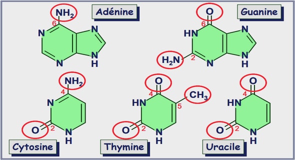
Active compounds identified 162 Compound 1 (Fig. 1) was isolated as an amorphous powder. 163 HRESI-TOF performed in the negative mode exhibited a deprotonated 164 molecular ion at m/z 108.0435 [M − H] 165 − indicating a molecular formula of C6H7NO (calcd. 108.0447) requiring 4° of unsaturation. 166 The 167 13C NMR spectrum revealed the presence of an oxygenated quaternary carbon at δ 151.2, another quaternary carbon at δ 117.4 168 and a methine carbon at δ 115.8. 169 The 170 1 H NMR spectrum of this small molecule displayed an intense signal of four aromatic protons at δ 6.49. As the spectra were realized 171 in CD3OD, the three remaining protons not observed as suggested the 172 molecular formula are exchangeable protons. Comparison with RMN 173 data of the sample indicated that compound 1 was a 4-aminophenol 174 (Sigma-Aldrich catalog). 175 The molecular formula of compound 2 (Fig. 1) was deduced as 176 C7H6O2 from the deprotonated molecular ion peak at m/z 122.0368 177 [M − H] 178 − observed in the HRESI-MS compatible with four degrees of unsaturation. Its 179 1 H NMR spectrum showed the presence of an aldehyde proton at δ 9.73 and two doublets at δ 6.9 (2H, J = 8.4 Hz, 180 H-3; H-5) and 7.8 (2H, J = 8.4 Hz, H-2; H-6). In comparison with 1, 181 the difference of 13 uma suggested that the amino group was replaced .